Anatomy Muscles Pelvis / Anterior Muscles Of The Pelvis / Muscles of the pelvic floor gross anatomy.. Muscles of the pelvic floor gross anatomy. There are many muscles that form the pelvic floor, including puborectalis, pubococcygeus, iliococcygeus and coccygeus. Ascending colon superior mesenteric vein superior mesenteric artery gonadal vessels linea semilunaris abdominal aorta linea alba inferior vena cava inferior mesenteric artery infe. Arcus tendineus levator ani and the ischial spine The muscles of the abdomen, lower back, and pelvis are separated from those of the chest by the muscular wall of the diaphragm, the critical breathing muscle.
The levator ani muscles consist of three. To support the abdominal and pelvic viscera The term pelvis is used to identify the area between the abdomen and the lower extremities. The muscles of the pelvis form its floor. The levator ani muscles are the largest group of muscles in the pelvis.
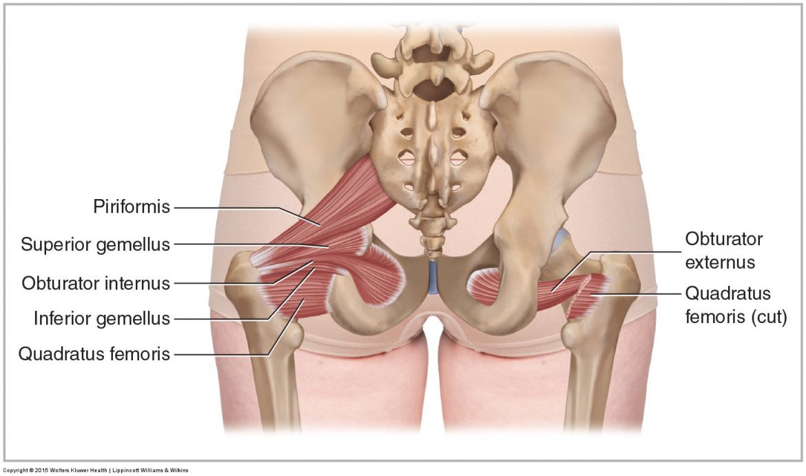
Muscles Of The Pelvis from learnmuscles.com The floor of the pelvis is made up of the muscles of the pelvis, which support its contents. These muscles can be grouped based upon their location and function. It can be divided into the greater pelvis and the lesser pelvis. These are the piriformis, obturator internus, obturator externus, gemellus superior, gemellus inferior, and quadratus femoris. The bony anatomy of the pelvic girdle consists of 3 bones and 3 joints. Muscles that attach from the pelvis to the trunk and cross the lumbosacral joint muscles that attach from the pelvis to the thigh/leg and cross the hip joint pelvic floor muscles that are located wholly within the pelvis Psoas consists of a pair of deep muscles (psoas major and iliacus) located on each side of the pelvis in the abdomen. Several muscles around the pelvis take part in movements of the thigh.
Psoas consists of a pair of deep muscles (psoas major and iliacus) located on each side of the pelvis in the abdomen.
These muscles have attachments to the pelvis as follows: This mri male pelvis axial cross sectional anatomy tool is absolutely free to use. The pelvis marks an important transition point between the thoracoabdominal region and the lower limbs. The pelvic floor muscles include; There are many muscles that form the pelvic floor, including puborectalis, pubococcygeus, iliococcygeus and coccygeus. Right posterolateral view of the lumbar spine. It is composed of three separate paired muscles; The pelvis forms the base of the spine as well as the socket of the hip joint. The medial surface provides attachment for both transverse perinei, obturator internus and externus, piriformis, coccygeus and levator ani muscles. Deep to the gluteus maximus, the piriformis, obturator internus, obturator externus. The levator ani muscles are the largest group of muscles in the pelvis. Use the mouse scroll wheel to move the images up and down alternatively use the tiny arrows (>>) on both side of the image to move the images.>>) on both side of the image to move the images. A proper kegel exercise means a full contraction and relaxation of the pc muscle.
The pelvis consists of the sacrum, the coccyx, the ischium, the ilium, and the pubis. Arcus tendineus levator ani and the ischial spine Deep to the gluteus maximus, the piriformis, obturator internus, obturator externus. These are the piriformis, obturator internus, obturator externus, gemellus superior, gemellus inferior, and quadratus femoris. The many muscles of the hip provide movement, strength, and stability to the hip joint and the bones of the hip and thigh.
Pelvic Floor Wikipedia from upload.wikimedia.org The pelvic floor muscles are comprised mainly of the levator ani muscles with somatic innervation from the lumbosacral plexus. There are many muscles that form the pelvic floor, including puborectalis, pubococcygeus, iliococcygeus and coccygeus. Some of the major pelvic muscles are as follows. The gluteal muscles are a group of three muscles named the gluteus maximus, the gluteus medius, and the gluteus minimus. The muscles of the pelvis and hip control the vast range of movement of the legs and torso. The pelvis's frame is made up of the bones of the pelvis, which connect the axial skeleton to the femurs, and therefore acts in weight bearing of the upper body. Deep to the gluteus maximus, the piriformis, obturator internus, obturator externus. Muscles an important group of muscles in the pelvis is the pelvic floor.
The tensor fascia latae is a thick, squarish muscle in the superior aspect of the lateral thigh.
These are the piriformis, obturator internus, obturator externus, gemellus superior, gemellus inferior, and quadratus femoris. Arcus tendineus levator ani and the ischial spine The bony anatomy of the pelvic girdle consists of 3 bones and 3 joints. The hip joint is one of the most flexible joints in the entire human body. The pelvis consists of the sacrum, the coccyx, the ischium, the ilium, and the pubis. It is composed of three separate paired muscles; Because of the lordotic curve and the thick musculature that overlies the lumbar spine, the only easily palpable bony landmark is the spinous process. Ascending colon superior mesenteric vein superior mesenteric artery gonadal vessels linea semilunaris abdominal aorta linea alba inferior vena cava inferior mesenteric artery infe. The function of the pelvic floor is to help assist with child birth, prevent incontinence and support organs within the pelvis. The pubococcygeus (pc) muscle is the muscle that runs the show in pelvic floor health. This mri male pelvis axial cross sectional anatomy tool is absolutely free to use. The pelvic floor muscles are comprised mainly of the levator ani muscles with somatic innervation from the lumbosacral plexus. Muscles that attach from the pelvis to the trunk and cross the lumbosacral joint muscles that attach from the pelvis to the thigh/leg and cross the hip joint pelvic floor muscles that are located wholly within the pelvis
Ascending colon superior mesenteric vein superior mesenteric artery gonadal vessels linea semilunaris abdominal aorta linea alba inferior vena cava inferior mesenteric artery infe. These muscles can be grouped based upon their location and function. In this section, learn more about the pelvic floor,. The tensor fascia latae is a thick, squarish muscle in the superior aspect of the lateral thigh. The pelvic floor muscles provide foundational support for the intestines and bladder.
1 from The pubococcygeus (pc) muscle is the muscle that runs the show in pelvic floor health. The four groups are the anterior group, the posterior group, adductor group. Find symptoms,causes and treatments of pelvic disease.for your health. The levator ani muscles are the largest group of muscles in the pelvis. Prominent bony landmarks are labeled. It can be divided into the greater pelvis and the lesser pelvis. It can be described as one of the bodies diaphragms. The function of the pelvic floor is to help assist with child birth, prevent incontinence and support organs within the pelvis.
The pelvis is the lower portion of the trunk, located between the abdomen and the lower limbs.
Deep to the gluteus maximus, the piriformis, obturator internus, obturator externus. It can be described as one of the bodies diaphragms. The pelvis marks an important transition point between the thoracoabdominal region and the lower limbs. The muscles of the pelvis form its floor. On the posterior side they are the glutei and on the anterior side the hip muscles extending into the thighs. The pelvis's frame is made up of the bones of the pelvis, which connect the axial skeleton to the femurs, and therefore acts in weight bearing of the upper body. It also helps stabilize the lateral aspect of the knee by pulling on the iliotibial tract (band), making it taut. The gluteal muscles are a group of three muscles named the gluteus maximus, the gluteus medius, and the gluteus minimus. The pelvic floor muscles are comprised mainly of the levator ani muscles with somatic innervation from the lumbosacral plexus. These are the piriformis, obturator internus, obturator externus, gemellus superior, gemellus inferior, and quadratus femoris. It acts as a synergist of the gluteus medius and iliopsoas in flexing and abducting the thigh. These muscles can be grouped based upon their location and function. The pelvis forms the base of the spine as well as the socket of the hip joint.

0 Comments:
Posting Komentar